| |
|
|
|
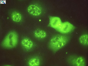
Immunocytochemical localization of splicing factors in rat liver cells showing cell cycle dependent alteration in their aggregate structure.
|
|
|
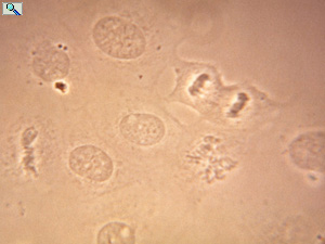
Phase contrast micrograph of rat liver cells shown in the immunocytochemical localization.
|
|
|
|
|
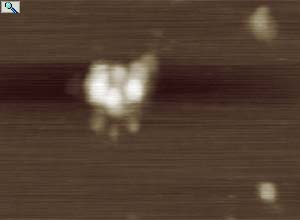
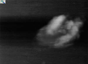
Atomic force Microscopy of affinity purified APOBEC-1 containing editosome assembled on the mooring sequence within a 498 nt segment of apoB RNA (data collected by the University of Vermont, core structure facility) The dimensions of the complex suggested a molecular mass of 650 kDa.
|
|
|
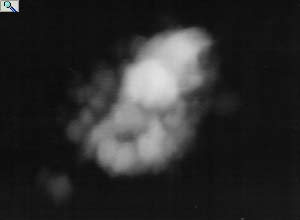
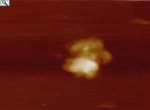
Another example of an affinity purified editosome.
|
|
|
|
|
Affinity purified editosome, RNase digested before analysis.
|
|
|
Another example of an RNase digested editosome.
|
|
|
|
|
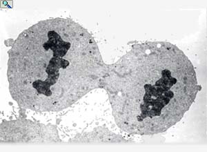
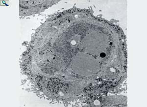
HeLa cell undergoing cell division (cytokinesis), grow in suspension culture.
|
|
|
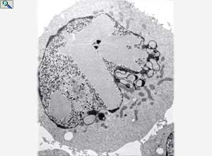
HeLa cell in late Adenovirus 5 infection showing viral particles within the nucleus and para crystalline arrays of viral proteins.
|
|
|
|
|
HeLa cell in late Adenovirus 5 infection showing disruption of internal membranes and release of viral particles to the cytoplasm.
|
|
|
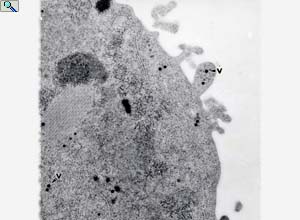
Periphery of HeLa cell in late Adenovirus 5 infection showing the release of viral particle (v) from a membrane bleb.
|
|
|

|
|
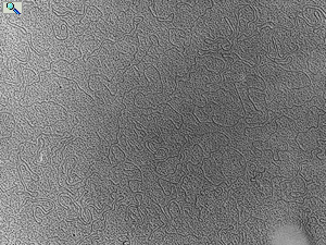
Electron micrograph of DNA fragments
|
|
|
High magnification electron micrograph of a rat liver thin section showing the distribution of ACF to chromatin in the nucleus (Nu), to the periphery of the endoplasmic reticulum (ER) in the cytoplasm but not within mitochondria (Mt).
|
|

|
|

)
)
)
)
)
)
)
)
)
)
)
)